
Introduction
I remember the first time I watched a freshly dissociated sample under the microscope — cells floating, some whole, some shredded — and I thought, we can do better. In routine protocols for tissue dissociation single cell prep, lab reports often show survival rates anywhere from 40% to 85% depending on method and operator skill. That wide spread (and yes, frustration) raises a clear question: which steps actually drive loss of viable cells, and how do we choose the right trade-offs between speed, yield, and phenotype preservation? I’ll walk you through what I’ve learned, with plain language and a few practical data points, so you can start testing smarter rather than hoping for better. Next, I’ll break down where standard approaches stumble and what that means for your downstream assays.
Part 1 — Why Many Protocols Fall Short: A Technical Look
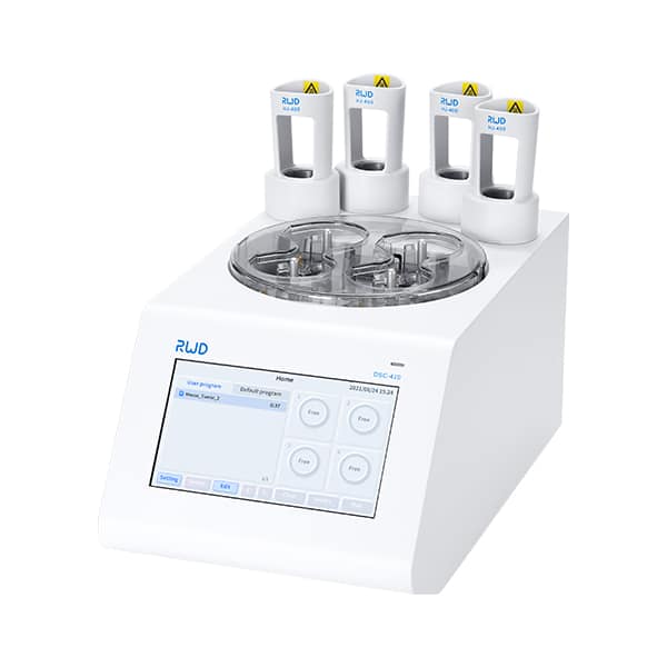
Let’s be direct: the tools and steps we inherit often contain hidden compromises. At the heart of the problem is the mismatch between biological material and mechanical or enzymatic treatment — and that’s where a modern tissue dissociator can change the game. Technically speaking, enzymatic digestion rates, mechanical shear, incubation times, and temperature control interact in non-linear ways. Shortcuts like over-agitation to speed throughput can fragment fragile cell types (neurons, endothelial cells) and skew cell-type proportions. Conversely, overly gentle handling preserves viability but yields clumped suspensions that confound cell sorting and single-cell RNA-seq library prep.
What’s the real bottleneck?
In simple terms: balancing cell viability with representative recovery. Enzymes (collagenase, dispase) break extracellular matrix but also stress cell membranes; mechanical agitation separates tissue but causes shear stress. Add microfluidics downstream and you suddenly care about droplet size, clog rates, and debris load. Look, it’s simpler than you think when you map each step to a measurable outcome — yield, viability, and transcript integrity. I’ve run side-by-side comparisons in-house: changing agitation pattern reduced doublets by nearly half but required a small trade in speed — funny how that works, right? These are the kinds of practical trade-offs you’ll want to measure before scaling.
Part 2 — Forward-Looking Principles and Practical Metrics
Moving ahead, I favor two complementary approaches: refine process controls and adopt smarter hardware. A well-tuned tissue dissociator gives repeatable shear profiles and controlled enzymatic mixing, which reduces operator-to-operator variability. In plain terms: automation doesn’t remove the need for judgement — it amplifies consistency. If you couple controlled digestion with staged filtration and a gentle wash protocol, you’ll see fewer dead cells and cleaner suspensions for flow cytometry or single-cell sequencing. I recommend testing one variable at a time: enzyme concentration, agitation frequency, and temperature ramp. Measure RNA integrity number (RIN), cell viability, and doublet rate after each change.
Real-world impact
I’ve watched teams shorten optimization cycles from months to weeks by adopting a modular workflow: (1) pre-processing to remove fat and connective tissue, (2) calibrated enzymatic digestion, and (3) mechanical dissociation tuned to tissue type. This reduces batch effects and improves reproducibility across experiments. You’ll still need judgment calls — tissue heterogeneity demands it — but the point is to make those calls data-driven. Also, consider downstream constraints: platforms like droplet microfluidics tolerate different debris loads than plate-based sorting. Plan accordingly — and yes, test small runs before committing large sample sets.
Closing — How to Evaluate and Choose a Protocol or Device
Weighing options can feel overwhelming. Here are three concrete metrics I use to evaluate any dissociation method or device: (1) Recovery Efficiency — percent of target cells recovered relative to input tissue mass; (2) Functional Viability — not just membrane integrity but functional readouts or RIN for transcriptomic work; and (3) Reproducibility — variance across operators and days. Use small pilot runs to collect these numbers and compare. If you prioritize single-cell RNA-seq, give extra weight to RNA quality and doublet rate. If you need surface markers for FACS, cell-surface antigen preservation matters most.

In short: choose approaches that let you measure trade-offs, then optimize those that matter for your assay. I’ve found that combining controlled enzymatic conditions with reproducible mechanical dissociation reduces downstream noise and saves time in the long run. — funny how that works, right? For practical adoption, check device specs for sample throughput, shear control, and compatibility with downstream platforms. If you want a starting point, consider vendors that provide clear performance data and real-world application notes. For more resources and product details, see BPLabLine.